Malaria Life Cycle

QUESTION

What is the life cycle of malaria?

ANSWER

Malaria is caused by a single celled organism in the genus Plasmodium. Five species of Plasmodium infect humans, but all follow a very similar life cycle, including two separate cycles of asexual reproduction in the human host (one in the liver, called the exo-erythrocytic cycle, and one in the blood, and specifically inside red blood cells, known as the erythrocytic cycle) and a sexual reproductive stage inside the mosquito definitive host (usually called the “vector”). A schematic of the full life cycle is below, courtesy of the CDC (www.cdc.gov).
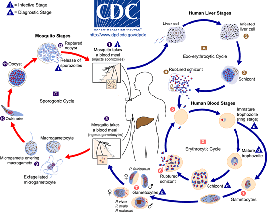
malaria life cycle CDC

Schematic of the malaria life cycle, courtesy of CDC (www.cdc.gov)

Three Stages of Malaria

QUESTION

What are the three stages of malaria?

ANSWER

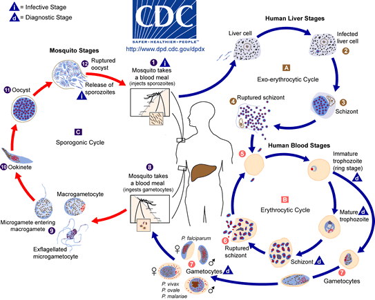
The three parts of the malaria life cycle are known as the exo-erythrocytic cycle, the erythrocytic cycle and the sporogonic cycle. The first two of these take place in the human (or other mammal) host, while the third occurs in the mosquito vector. The below diagram, courtesy of the CDC, shows the complete life cycle of malaria in more detail.

malaria life cycle CDC

Schematic of the malaria life cycle, courtesy of the CDC (www.cdc.gov)

Schizont

QUESTION

What is a schizont?

ANSWER

A schizont is a malaria parasite which has matured and contains many merozoites, which are the parasite stage that infects red blood cells.

Schizonts can be produces during two separate phases of the life cycle within the human host: first in the hepatocytic cells in the liver (when sporozoites mature) during the exo-erythrocytic cycle and then within the red blood cells during the erythrocytic cycle (when trophozoites mature and divide).

When malaria parasites do not immediately mature into schizonts in the liver (as can be the case with Plasmodium vivax and P. ovale infections), the parasite instead becomes a hypnozoite, which can lay dormant in the liver for many weeks or even months (or, in rare case, years), and produce relapse of infection at a much later date.

Pathophysiology of Malaria

QUESTION

What is the pathophysiology of malaria?

ANSWER

Malaria causes disease through a number of pathways, which depend to a certain extent on the species. Malaria is caused by a single-celled parasite of the genus Plasmodium; there are five species which infect humans, being Plasmodium falciparum, P. vivax, P. ovale, P. malariae and P. knowlesi.

All these species are introduced into the human blood stream through the bite of an infected mosquito; the life stage of malaria at this point is called a “sporozoite”, and they pass first to the liver, where they undergo an initial stage of replication (called “exo-erythrocytic replication”), before passing back into the blood and invading red blood cells (called “erythrocytes”, hence this is the “erythrocytic” part of the cycle). The malaria parasites that invade red blood cells are known as merozoites, and within the cell they replicate again, bursting out once they have completed a set number of divisions. It is this periodic rupturing of the red blood cells that causes most of the symptoms associated with malaria, as the host’s immune system responds to the waste products produced by the malaria parasites and the debris from the destroyed red blood cells. Different species of malaria rupture the red blood cells at different intervals, which leads to the diagnostic cycles of fever which characterise malaria; P. vivax, for example, tends to produce cycles of fever every two days, whereas P. malaria produces fever every three.

In addition, Plasmodium falciparum produces unique pathological effects, due to its manipulation of the host’s physiology. When it infects red blood cells, it makes them stick to the walls of tiny blood vessels deep within major organs, such as the kidneys, lungs, heart and brain. This is called “sequestration”, and results in reduced blood flow to these organs, causing the severe clinical symptoms associated with this infection, such as cerebral malaria.

More details on the exact biochemical mechanisms for sequestration and its effect on the pathology of the infection can be found on the Tulane University website.